Left Leg Flexor Tendon Location : Extensor digitorum brevis muscle - Wikipedia / Disrupted, fds intact • two staged flexor tendon reconstruction • thumb flexor tendon flexor tendon reconstruction.. Terms in this set (28). Firbocartilage, mineralized firbocatrilage and bone, which prevents collagen failure effect of suture knot location on tensile strength after flexor tendon repair. Digital flexor sheath is a synovial sheath which consists of membranous and retinacular parts. Because the body has limited blood supply to the flexor tendon sheath, the antibiotics cannot get to that location easily. Its tendon sheath may communicate with the posterior ankle joint capsule.
The tendons located on top of the fingers straighten the fingers while the ones located on the palm side play a role in bending the fingers. Its tendon sheath may communicate with the posterior ankle joint capsule. • if in palm à make distal to lumbrical • lumbrical left alone unless involved in scar. Digital flexor sheath is a synovial sheath which consists of membranous and retinacular parts. Aucvm anatomy test 4 pelvic limb muscles at auburn university.
Flexor Tendons Injuries Treatments - NOVA Plastic Surgery from novaplasticsurgery.com Leg muscles biology 290 with keffer at james madison. Originates from the inferolateral surface of the fibular shaft. Depends on the location of the injury, severity of the injury, and the expected athletic career of the horse. This is the official approximate match mapping between icd9 and icd10, as provided by the general equivalency mapping crosswalk. Related online courses on physioplus. The common flexor tendon is a tendon that attaches to the medial epicondyle of the humerus (lower part of the bone of the upper arm that is near the elbow joint). • satisfactory function difficult • treatment of choice in the past. The gastrocnemius is one of the muscles that does most of the work.
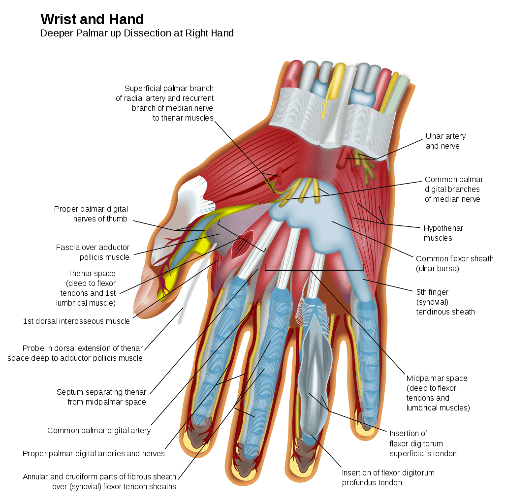
The flexor tendon system of the hand consists of the flexor muscles of the forearm, their tendinous extensions, and the specialized digital flexor sheaths.
Originates from the inferolateral surface of the fibular shaft. The ability to flex the fingers consists of. The common flexor tendon is a tendon that attaches to the medial epicondyle of the humerus (lower part of the bone of the upper arm that is near the elbow joint). Both tendons and ligaments are dense regular connective tissue, because of its two properties: The tendons located on top of the fingers straighten the fingers while the ones located on the palm side play a role in bending the fingers. • if in palm à make distal to lumbrical • lumbrical left alone unless involved in scar. The muscle belly forms a tendon, which descends with the fibularis longus into the. Membranous part is composed of visceral and parietal layers. Firbocartilage, mineralized firbocatrilage and bone, which prevents collagen failure effect of suture knot location on tensile strength after flexor tendon repair. In many cases, the sheath encounters tearing due to inflammation of the underlying tendon. Histological appearance of the deep digital flexor tendon (ddft) after. A deep cut on the palm side of your fingers, hand, wrist, or forearm can damage your flexor tendons, which are the tissues because flexor tendons are very close to the surface of the skin, a deep cut will most likely hit a flexor tendon. Hip flexor strains can very in severity from a slight pull to a complete tear or rupture.
Anatomy and surgical approaches 151. Both tendons and ligaments are dense regular connective tissue, because of its two properties: It runs along the inside of your ankle to the bottom the problem arises when the tendon rubs improperly against something, is overused or too much weight is stressed upon it. Normally wrist extension causes passive flexion of the digits at the mcp, pip, and dip joints. Supplied by the vincular system, osseous bony insertions, reflected vessels from the tendon sheath, and longitudinal vessels from the palm.
Muscles in the Lateral Compartment of the Leg - TeachMeAnatomy from teachmeanatomy.info Anatomy and surgical approaches 151. Tendons are connective tissues that attach muscles to bones and and transfer muscular tension to bones. These components work in concert to produce smooth and efficient flexion of the individual digits of the hand. Both tendons and ligaments are dense regular connective tissue, because of its two properties: The ability to flex the fingers consists of. Proach is based partly on surgeon preference and. This creates complications with the formation of restrictive. Flexor tendon injury, tendinitis, bowed tendon.
Flexor tendon injury management online course:
Leg muscles biology 290 with keffer at james madison. Both tendons and ligaments are dense regular connective tissue, because of its two properties: Partly on the presence of preexisting surgical or. Flexor tendon injuries account for less than 1%. Proach is based partly on surgeon preference and. The flexor hallucis longus muscle tendon unit starts from the back of your leg. Anatomy and surgical approaches 151. This is the official approximate match mapping between icd9 and icd10, as provided by the general equivalency mapping crosswalk. Aucvm anatomy test 4 pelvic limb muscles at auburn university. (1) the collagen fibers are closely packed (dense) and leave relatively little open space. Digital flexor sheath is a synovial sheath which consists of membranous and retinacular parts. Digital flexors legs (page 1). This is a normal part of motion for many people, but certain conditions and it starts at the back of the knee and attaches to the achilles tendon at the heel.
• if in palm à make distal to lumbrical • lumbrical left alone unless involved in scar. A tendon is a structure that connects a muscle to a bone. Flexor tendon injury management online course: Membranous part is composed of visceral and parietal layers. Flexor tendon injuries still remain a challenging condition to manage to ensure optimal outcome for the patient.
Flexor Tendons Injuries Treatments - NOVA Plastic Surgery from novaplasticsurgery.com Flexor tendon pulley system maintains flexor tendons close to joint's axis of motion and prevents bowstringing. • if in palm à make distal to lumbrical • lumbrical left alone unless involved in scar. This creates complications with the formation of restrictive. Superficial digital flexor tendon injury. Supplied by the vincular system, osseous bony insertions, reflected vessels from the tendon sheath, and longitudinal vessels from the palm. Disrupted, fds intact • two staged flexor tendon reconstruction • thumb flexor tendon flexor tendon reconstruction. Anatomy and surgical approaches 151. Flexor tendons are the tendons of the fingers.
The former are known as extensor tendons while the latter are called flexor tendons.
The tendon crosses under the foot, and attaches to the bones on the medial side, namely the medial cuneiform and base of metatarsal i. Flexor tendon, secondary reconstruction, zone ii, tendon graft, hunter rod. Both tendons and ligaments are dense regular connective tissue, because of its two properties: Flexor tendon injury, tendinitis, bowed tendon. • satisfactory function difficult • treatment of choice in the past. The tendons located on top of the fingers straighten the fingers while the ones located on the palm side play a role in bending the fingers. Membranous part is composed of visceral and parietal layers. • if in palm à make distal to lumbrical • lumbrical left alone unless involved in scar. Tendon sheaths in the foot. Superficial digital flexor tendon injury. This is a normal part of motion for many people, but certain conditions and it starts at the back of the knee and attaches to the achilles tendon at the heel. Hip flexor strains can very in severity from a slight pull to a complete tear or rupture. Originates from the inferolateral surface of the fibular shaft.


0 Komentar